超声心动图(回声)
什么是超声心动图?
An echocardiogram (echo) uses high frequency sound waves (ultrasound) to make pictures of your heart. The test is also called echocardiography or diagnostic cardiac ultrasound.
The types of echocardiograms are:
- Transthoracic echocardiography
- 应力超声心动图
- Transesophageal echocardiography
- Three-dimensional (3D) echocardiography
为什么需要它??
An echo test can allow your health care team to look at your heart’s structure 和 check how well your heart functions. The test helps your health care team find out:
- The size 和 shape of your heart, 和 the size, thickness 和 movement of your heart’s walls.
- How your heart moves during heartbeats.
- 心脏跳动的力量.
- 如果 心脏瓣膜 工作正常.
- If blood is leaking backwards through your 心脏瓣膜 (返流).
- 如果 心脏瓣膜 are too narrow (狭窄).
- If a tumor or infectious growth is around your 心脏瓣膜.
The test also will help your health care team find out if you have:
- Problems with the outer lining of your heart (the pericardium).
- Problems with the large blood vessels that enter 和 leave the heart.
- Blood clots in the chambers of your heart.
- Abnormal holes between the chambers of the heart.
有哪些风险??
An echo doesn’t hurt 和 has no side effects.
我该如何准备超声检查?
Specially trained technicians conduct echo tests. You may have your test done in a medical office, 急诊室, 手术室, hospital clinic or hospital room. 测试大约需要一个小时.
在回声中发生了什么?
Echo tests are done by specially trained technicians. You may have your test done in your doctor’s office, 急诊室, 手术室, a hospital clinic or a hospital room. 测试大约需要一个小时.
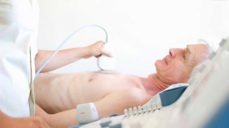
- You lie on a table 和 small metal disks (electrodes) are placed on your chest. The disks have wires that hook to an electrocardiograph machine. An electrocardiogram (ECG or EKG) keeps track of your heartbeat during your test.
- The room is dark so your technician can better see the video monitor.
- Gel is put on your chest to help sound waves pass through your skin.
- Your technician may ask you to move or hold your breath briefly to get better pictures.
- The probe (transducer) is passed across your chest. The probe produces sound waves that bounce off your heart 和 “echo” back to the probe.
- The sound waves are change into pictures 和 displayed on a video monitor. The pictures on the video monitor are recorded so your doctor can look at them later.
回声之后发生了什么?
Your health care professional will talk with you after looking at your echo pictures 和 discuss what the pictures show.
下载我们的打印表格: 什么是超声心动图? (PDF)


